Chalk Talk: Exploring the biological processes that underlie Cascade by TinkRNG
Summary: A deep dive into the exciting biology represented in Cascade by TinkRNG, and a color palette reveal.
The patterns that underlie every aspect of life are as beautiful as they are complex, and mankind’s pursuit to understand them and manipulate them are just as enthralling. At TinkRNG, we are inspired by biology and biological engineering. One way we are sharing this passion with the world is through the unique lens of generative art. In our white paper describing our first project in this space, Cascade by TinkRNG, we introduced our novel approach for integrating biological information with generative art. Here, we take a step back to feature the exciting biology represented in our first collection. Our goal is to convey biological concepts to the general public in an inspiring and informative way. Think of this like a “chalk talk,” where we are sharing some cool ideas with you. (This format is embodied by, if nothing else, the hand-drawn sketches that are featured in this article.)
At the conclusion of this article, as a reward for your attendance, we reveal for the first time the bio-inspired color palettes of Cascade. By the end, we hope that you will connect with the art, and the message and meaning behind it, as we do.
Recall: Cascade is a framework to represent proteins using generative art
As previously discussed in our first article, the general premise of Cascade is to represent patterns in the human proteome (i.e. the set of proteins encoded in the human genome and expressed in human cells) using generative art. Each piece represents a unique protein, with properties derived from information including the protein’s amino acid sequence, the cellular compartment where the protein localizes to, and the biological process that the protein is involved in. The framework we developed is summarized with the example art construction shown in Fig 1. The vector field is inspired from the biological process, the shape style is inspired from the cellular compartment, and the number and color of shapes are derived from the actual amino acid sequence of the protein.

For a complete description of this process, please reference our official white paper for the Cascade project. With this refresher out of the way, let’s dive head-first into some of the exciting biology behind the art!
Cell Migration: A persistent random walk
We’ll start with some of the biological processes represented in Cascade, which are represented by the vector fields in the art. These are based on the biochemical and physical concepts behind the biological processes, as well as the exciting experimental approaches used to study them. One of these processes is cell migration, i.e. how cells move around. During the development of a human embryo, cells migrate to form the organized tissues and organs of the body. Additionally, when a tissue is damaged (on our skin or even on organs inside the body), cells must migrate to the site to protect against infection and repair the tissue. In addition to its normal functions, cell migration can also go awry, leading to the progression of diseases, such as the invasion of cancer cells. For all these reasons, and many more, understanding how cells migrate has been a major area of biological research and continues to be.

Many of the various types of cells in the human body undergo migration at some point during the human lifespan. The migration of each cell type has unique features and mechanisms, but there are some general principles that apply more broadly. One form of migration employed by a variety of cells involves adhering, or gripping, onto a surface such as the extracellular matrix or other cells. During this type of migration, cells move by sticking to and pulling themselves across the surface, similar to crawling. This happens in a cycle. Cells first become polarized in a specific direction, which means that the front of the cell and the rear of the cell adopt different compositions and shapes (Fig 2A). At the front, the cell extends a protrusion, such as a lamellipodium, which it uses to extend itself along the surface and explore its environment. Here, cells form adhesions, essentially “sticky patches,” that mechanically connect the cell to the extracellular environment (substrate). These serve as a grip, which allows the cell to contract and translocate forward. Lastly, it must disassemble adhesions at the rear and retract its rear. Migration proceeds through cycles like this. However, the cell is not perfect at steering itself. Even when there is an external signal it is following, its direction wobbles over time, due partly to noise in the signal and how it is sensed (more on this later). When the motion of a cell is tracked over time, as done in experiments watching the movement of cells in two dimensions along an artificial surface using a microscope (Fig 2B), the trajectory of the cell appears as bursts of linear movement in one direction followed by a change in direction and then another cycle of movement in the new direction. Without an external signal, this movement pattern resembles a persistent random walk (Fig 2C). In this case, on average, the cell does not move away from its starting point. In contrast, when there is an external signal to guide the cell, it moves in a biased manner (still with some amount of randomness) toward the direction of the signal (Fig 2D). One type of signal commonly used to guide cell migration, naturally in the human body and artificially in the laboratory, is a spatial gradient of a chemical that the cell can sense. Migration along this gradient is referred to as chemotaxis. Looking at the artwork, the vector field used to represent the biological process of Cell Migration in Cascade (Fig 2E) is inspired from the appearance of these cell trajectories!
Signal Transduction: Circuits for biological systems
To sense and respond to its environment, a cell must recognize stimuli at the cell surface and convert them into signals that can be interpreted and relayed in order to initiate a response. This includes controlling fast, dynamic events like the generation of protrusions or contractile forces involved in migration described in the last section, as well as the regulation of slower processes like cell growth or differentiation through changes in the expression of genes. Signal transduction underlies nearly every cellular process and can be targeted with chemicals to treat certain diseases. As the basis for information transfer inside cells, this also provides a toolkit that researchers are currently using to engineer synthetic cells to perform new tasks. In fact, the name of our first generative art project, Cascade, actually refers to design principle found in cell signaling circuits, described later in this article!

Signal transduction occurs in pathways, which are networks that involve specific proteins and serve specific purposes. However, despite the high level of diversity and complexity found in cell signaling pathways, there is a common toolkit that evolution has seemingly used to assemble many of these pathways. First, an external signal, such as a molecule secreted by another cell or tissue is detected at the cell surface by specific receptor proteins with pockets that are designed to bind specifically to the molecule (Fig 3A). This event kicks off many signal pathways. But how is this receptor-binding event relayed inside the cell? Recall that proteins have specific three-dimensional structures and that these structures dictate their functions. For instance, a protein that catalyzes a reaction has a special pocket structure where it binds reactants and facilitates their conversion into products. So, back to the receptor protein. These proteins typically span the cell membrane, with a part outside the cell and another part inside the cell. The external binding event can be relayed inside the cell if the binding induces a change in the structure of the protein, especially the part on the inside of the cell. This enables the binding of a messenger protein from the cytosol inside the cell, which means information has now been transferred from the outside of the cell to the inside of the cell. Typically, the signal is propagated by these messengers inside the cell by a series of events in which one protein binds another protein and modifies its structure and thus function. If it turns on a function, then we say it “activated” the protein. If it turns off a function, then we say it “inhibited” the protein. These interactions, which happen as a chain or sequence of events, form the basis of signal transduction. They are often referred to as signaling cascades (the namesake for our first project). Downstream of these cascades, activated proteins perform functions that are involved in generating responses. For example, the activation of proteins that remodel the cytoskeleton might be involved in changing the speed or direction of a migrating cell. Often, these signaling cascades end in the nucleus with transcription factors, which change how the proteins encoded in the human genome are expressed. Overall, these signaling pathways can be represented with control diagrams (Fig 3B) that indicate the components (proteins) and how they interact (activation, inhibition, etc). When downstream proteins affect the function of upstream proteins, this is called feedback. Positive feedback (activation) serves to enhance the response and negative feedback (inhibition) serves to stop the response. Together with other interactions, these control mechanisms allow the cell to respond precisely to its external environment. Overall, the complex interaction networks that constitute cell signaling pathways inspire the vector field used to represent this biological process in Cascade (Fig 3C).
Cell Division and Cell Adhesion: Drawing inspiration from the physical shapes of cellular machines
The first two biological processes we discussed, cell migration and signal transduction, were represented with vector fields inspired by our understanding of how the biological processes work. Next, we turn our attention to a different design concept, where we use the physical shapes and structures that execute the cellular processes to inform the vector fields. To replicate, cells duplicate their genomic information (chromosomes) and then divide. During this process of (mitotic) cell division, duplicated chromosomes must be separated and partitioned into two daughter cells. To do this, the cell forms a structure known as the mitotic spindle, which is composed of two poles (centrosomes) and an array of spindles (microtubules) extending from one pole to the other (Fig 4A). During mitosis, duplicated chromosomes align along the center of the cell and are attached to these spindles, which are used to physically separate the chromosomes into the two resulting daughter cells. The vector field for the cell division process resembles the mitotic spindle.
Similarly, the vector field for cell adhesion is inspired by the structures involved in the process. In the body, most cells stick to a special matrix, called an extracellular matrix, which is a fibrous network of proteins. Cells stick to the matrix to become anchored and positioned with respect to other cells in a tissue. This is critical for tissue structure and function. These adherent cell types are initially rounded when they come in contact with an extracellular matrix, after which they attach to the matrix through specialized adhesion structures, which are composed of adhesion receptor proteins that bind complementary proteins in the extracellular matrix. The vector field for cell adhesion is inspired by the rounded shape of a cell upon initial contact with the fibrous extracellular matrix (Fig 4B).

Cell Compartments: A brief tour
As previously discussed, in addition to inspiring the art’s vector field from the biological process that the protein is involved in, we also inspire the bar shape from the location in which the protein is primarily found!
The cell is divided into compartments where specific proteins are located and specific jobs are carried out. In the white paper for Cascade we outlined in detail how the geometrical properties of these cellular compartments inform the artistic shapes used for a protein and thus encodes the protein’s cellular localization. Here, we take a closer look at a few of these cellular compartments to highlight how the biochemical and biophysical structures connect to the artistic depictions.
The plasma membrane, which surrounds and encloses the cell, is formed by a lipid bilayer (Fig 5A). The lipid bilayer is formed by units called phospholipids, which have a hydrophilic head and hydrophobic tails. Due to the chemical properties of these molecules, in water they can self-assemble into a bilayer structure with the phosphate heads facing toward the solution and the hydrophobic tails shielded together. The artistic depiction of proteins that localize to the plasma membrane is informed by the appearance of the lipid bilayer. Furthermore, the main compartment of the cell, called the cytosol, houses many of the signaling pathways described earlier. Despite the usual representation of the cytosol as an empty space, it is actually packed with an abundance of proteins, which are like little globs bumping into each other (Fig 5B). The artistic depiction of proteins that are located in the cytosol is informed by the appearance of these globular proteins. Turning to the cytoskeleton, it is composed of long filaments that provide structural integrity to the cell. One type of filament, actin, forms many of the dynamic structures the cell uses to move and change its shape. In these actin networks, filaments composed of actin monomers are constantly polymerized and depolymerized, and they are also linked together by special proteins that connect them to form more ordered structures (Fig 5C). Thus, the long cytoskeletal filaments inspire the shapes used to represent cytoskeletal proteins in Cascade. Lastly, the nucleus houses the genetic information of the cell, in the form of DNA. This type of DNA is double-stranded and forms a helix structure, which inspires the shape used to represent proteins that localize to the nucleus (Fig 5D).

Cascade Color Palette Reveal
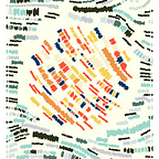
As a reward for attending our “chalk talk,” we now reveal for the first time the Cascade color palettes. In total there are 16 palettes, each with a frequency (rarity in the collection) and a biologically inspired name. For more information on how the color palette is used to depict the sequence of amino acids in a protein, we direct you back to the official white paper. Each color palette was named after a term used in molecular or cellular biology, which is both central to the Cascade project, or microscopy, which is a major tool used to visualize and study these processes. Here, we highlight a few of our favorite names, but we encourage readers to seek out more information about the terms used in the names of our color palettes!
We start with the most common color palette, which is the signature palette for our first collection and shares its name, “Cascade” (Fig 6). Along with this palette, there are four more palettes that are common in the collection, occurring each with 10% frequency (Fig 7). Two of these palettes, “Agonist” and “Antagonist,” have names that reflect their opposing natures. An agonist is a molecule that binds a cell receptor and activates a downstream pathway (Fig 8A), while an antagonist is a molecule that binds a receptor, blocking the agonist from binding and preventing the activation of the downstream pathway. In the next tier of color palettes (Fig 9), which occur with 8% frequency, “Trypan Blue” is named after a substance commonly used to distinguish living and dead cells under the microscope. This dye permeates the membrane of dead but not living cells (Fig 8B). Next, there are five color palettes that each occur with 4% frequency (Fig 10). “Dark-field” and “Bright-field” are inspired by forms of transmitted light microscopy, a technique that was instrumental in the characterization of cell migration described earlier in this article. Lastly, there are the ultra-rare (2% frequency, Fig 11) and unique (1% frequency, Fig 12) color palettes. A personal favorite is the name “Bandpass 434–17 nm” used for the monochromatic blue color palette. Some explanation about this name. Fluorescent dyes and proteins are commonly used to label proteins and visualize them inside cells. Fluorescent molecules have characteristic spectra corresponding to the wavelengths of light needed to excite them and the wavelengths of light they emit after being excited (Fig 8C). To visualize these molecular tags on a fluorescence microscope, special filters are used to selectively transmit certain wavelengths of light. Often these filters are “bandpass,” meaning they pass a narrow band of wavelengths. For exciting a blue fluorophore, such as a Cyan Fluorescent Protein (CFP), one might use a bandpass filter with a center wavelength of 434 nm and a width of 17 nm, which is depicted in Fig 8C and serves as the name for our monochromatic blue color palette!


Citations
No citations for this article.
Note: For citations and attributions related to the design and implementation of the Cascade project, including the sources for biological databases, which is not a topic covered in this article, see the official white paper: https://medium.com/@tinkrng/cascade-by-tinkrng-6f20bccfbf1d
***
Disclaimer: The UniProt Consortium and The Gene Ontology Consortium do not endorse or sponsor projects by TinkRNG, including Cascade by TinkRNG or Cascade Genesis by TinkRNG. TinkRNG is not granted any official status by The UniProt Consortium and The Gene Ontology Consortium. Furthermore, Digital Art from TinkRNG is intended solely to be artwork. TinkRNG does not modify or distribute any scientific data, and Cascade by TinkRNG and Cascade Genesis by TinkRNG are not intended for any scientific or other use. Ownership of Digital Art from TinkRNG does not include any of the biological information associated with artistic decisions used to create the art.
Terms of Use Agreement for Cascade by TinkRNG and Cascade Genesis by TinkRNG: https://www.tinkrng.io/termsofuse.pdf.